Multimodal Brain Modeling
Coordinator: François-René Molino
Our research focuses on the analysis and modeling of data from various magnetic resonance imaging (MRI) modalities, both structural and functional, to develop new model-based clinical strategies.
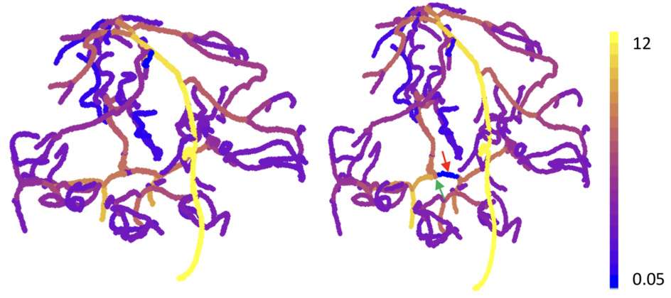
Our most significant research area in recent years has been the modeling of intracranial blood flow from multimodal imaging, providing an individualized description for each patient. Our team has developed a comprehensive environment for extracting multimodal structural data and input/output flow data, which enables the generation of intracranial flow simulations. The model description relies on decomposing the circulatory system into compartments defined from the structural data. This has the advantage of reducing the system to a set of ordinary differential equations, the integration of which can be made highly efficient.
Clinically, another important part of our work is dedicated to studying the evolution over time of low-grade gliomas, which are slow-growing brain tumors, in order to improve their monitoring and therapeutic management thanks to the ability to predict their progression early on. This area is currently under development. One of the key objectives of this work will be to compare the predictive power of methods based on physical modeling (growth models, the most common of which are reaction/diffusion models) with the rapidly expanding methods based on deep learning.
All of these activities reflect the current state of a collaboration established some fifteen years ago between Theoretical Physics and the Gui de Chauliac University Hospital, more specifically the I2FH platform (Institute of Functional and Human Imaging https://www.chu-montpellier.fr/fr/plateformes-recherche/imagerie-
fonctionnelle-i2fh). Concerning the I2FH platform, the people involved are Nicolas Menjot de Champfleur (PU-PH), Emmanuelle Le Bars (IR), and Jeremy Deverdun (IE).