Rechercher
Accueil > La Recherche > Axes & Equipes > Matière Molle & Verres > Equipe : Matière Molle > Thème : Physique de systèmes biologiques et biomimétiques
DIGITAL HOLOGRAPHIC MICROSCOPY FOR BIO IMAGING
publié le , mis à jour le
DIGITAL HOLOGRAPHIC MICROSCOPY FOR BIO IMAGING
People involved : Daniel Alexandre (CR), Alexey Brodoline (PhD), Dario Donnarumma (Postdoc), Michel Gross (DR), Nicolas Verrier (PostDoc)
1. Laser Doppler holographic microscopy in transmission : application to fish embryo imaging.
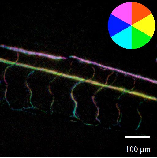
We have extended Laser Doppler holographic microscopy to transmission geometry. The technique is validated with living fish embryos imaged by a modified upright bio-microscope. By varying the frequency of the holographic reference beam, and the combination of frames used to calculate the hologram, multimodal imaging has been performed. Doppler images of the blood vessels for different Doppler shifts, images where the flow direction is coded in RGB colors or movies showing blood cells individual motion have been obtained as well. The ability to select the Fourier space zone that is used to calculate the signal, makes the method quantitative.

Fig. Image of the blood vessels of a zebrafish embryo that show with colors the direction of the RBCs flux.
2. 4D (x,y,z,t) holographic microscopy of zebrafish larvae microcirculation with double illumination [2]
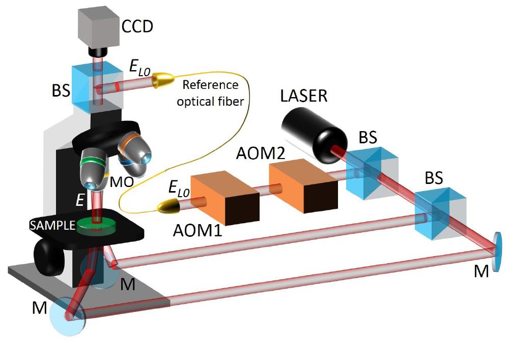
An original technique that combines digital holography, dual illumination of the sample and cleaning algorithm 3D reconstruction has been developped. It uses a standard transmission microscopy setup coupled with a digital holography detection. The technique is 4D, since it allows to determine, at each time step, the 3D locations (x ;y ;z) of many moving objects that scatter the dual illumination beam. The technique has been validated by imaging the microcirculation of blood in a fish larvae sample (the moving objects are thus red blood cells RBCs). Videos showing in 4D the moving RBCs superimposed with the perfused blood vessels are obtained.
Fig. Double illumination setup usedto perform 4D holography. AOM1, AOM2 : acousto-optic modulators (Bragg cells) that shift the frequency of the local oscillator beam ; MO : microscope objective ; M : mirror ; BS : angularly tilted cube beam splitter. E and ELO : signal and reference complex fields.
References
- [1] Verrier, N., Alexandre, D., & Gross, M. (2014). Laser Doppler holographic microscopy in transmission : application to fish embryo imaging. Optics express, 22(8), 9368-9379. https://arxiv.org/abs/1404.7781
- [2] D. Donnarumma, A. Brodoline, D. Alexandre, and M. Gross. 4d holographic microscopy of zebrash larvae microcirculation. Optic Express 24(3) : 26887-26900, 2016 https://hal.archives-ouvertes.fr/hal-01363227/


