Rechercher


Accueil > La Recherche > Axes & Equipes > Axes transverses > Graphène
Raman spectroscopy of graphene exfoliated on SiO2 /Si
publié le
To this aim, our expertise in studying vibrational properties of carbon nanotubes was recently extended to graphene. In particular, we use the Raman spectroscopy and Atomic Force Microscopy (AFM) to characterize graphene samples and investigate the effect of stacking (Bernal and/or misoriented) on the electronic band structure.
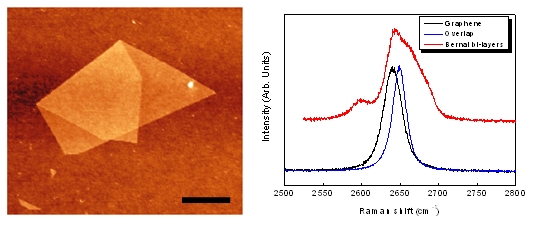
The samples are prepared by mechanical exfoliation of graphite and transferred to Si/SiO2 substrate. Naturally, some graphene flakes are folded or overlapped, as showed in Figure 1a. In this configuration, AFM is used to measure the number of graphene layers. Figure 1b compares the second order 2D peak of graphene, overlapped (misoriented) bi-layer graphene and Bernal bi-layer graphene. The unique 2D peak for a misoriented graphene bi-layer compares well with graphene signature although its frequency is different. Moreover, the Raman fingerprint of the AB stacked Bernal (oriented) bi-layer is significantly different with respect to the others systems. In agreement with theoretical calculations, these measurements indicate that the interaction between layers in the misoriented sample is weak and doesn’t result in an electronic band splitting contrary to the case of a Bernal bi-layer for which four components are observed.
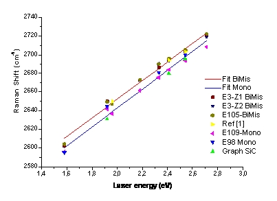
To go further in the understanding of the electronic structure and the phonon dispersion curves a current project is devoted to determine the dispersion of the 2D peak with the laser excitation energy (Figure 2). For instance, monolayer and misoriented bi-layer samples display parallel dispersion indicating similar Fermi velocity for both systems. The measurement of samples on other substrates (like epitaxial graphene on SiC) is also in progress.